Welcome to the Focal Point

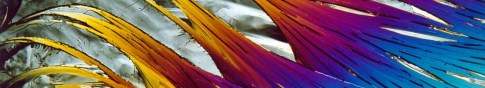
The Focal Point is the point at which rays of light come together to form an image; the point at which things become clearer or in focus.  In life it can also be the center of attention or purpose.  Focal Point Mineralogy, LLC was created by Julian Gray, a professional geologist specializing in mineralogy.  Based in of Hillsboro, Oregon, Julian created this site to be a Focal Point through which to share useful resources for mineralogy, photography, micro-analysis of minerals, and tips for mineral collecting, exhibition, and curation.  It also serves to highlight services that Julian provides such as lectures, educational outreach, consultation, and photography.

This home page serves as a blog with interesting articles including (beginning April 3, 2023) #MicroscopyMonday post.  Throughout the site you will find articles and links to resources and beautiful images illustrating the art of science.  Follow Focal Point Mineralogy on Facebook, Twitter (@FPMinerals), and Instagram (FPMinerals).

The contributions of James Dwight Dana & Edward Salisbury Dana to systematic mineralogy

Dana’s System of Mineralogy

Mineralogy took tremendous leaps forward in the nineteenth and early twentieth century, the science of thanks to the landmark contributions of James Dwight (1813-1895) and his son, Edward Salisbury Dana (1849-1935), both Yale geology professors. Although not the first mineralogy references or attempts at classification, the publication of these text on systematic mineralogy are remarkable.

Continue reading

How was this image made?

Thin section of dunite in crossed polars
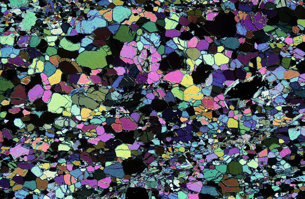
First off, what is this?  This is a photomicrograph of dunite, an ultramafic rock composed almost entirely of the mineral olivine.  The vivid colors were produced by viewing the subject, a thin section of the rock containing birefringent crystals, through crossed polarizing filters. Continue reading

What is this image?

Myristic acid at 100x viewed using cross polarized light

The striking image currently on the website banner and on my business card is a photo taken through a microscope.  The subject is myristic acid, an organic compound derived from Myristica fragrans, better known as nutmeg.  Myristic acid is found in many other plant oils.  To make the microscope slide, a few grains of the myristic acid, a white powder, were placed on a microscope slide and heated.  Myristic acid has a low melting temperature and crystallizes quickly when the heat is removed returning it to its original white crystalline solid state.  But each time it recrystallizes it produces a different whimsical microscopical world of interlocking patterns.  The image was magnified 100x and viewed through polarizing filters to reveal these fantastic colors, called “interference colors”. 

This image won best overall image in the Photomicrography Competition at the 2013 Inter/Micro, an International Microscopy Symposium held annually at the McCrone Research Institute in Chicago, Illinois.  The image was then featured on a cover of The Microscope and the program for Inter/Micro 2014.

Photos of magazine covers featuring photographs by Julian Gray

Stacked images: how are they made?

Recent developments in photography have lead to a flood of great images with tremendous sharpness or resolution.  Here’s an example photograph of a spherical cluster of blades of vauxite crystals from the Siglo Veinte mine, Llallagua, Potosi, Bolivia.  The ball of crystals is 1 mm wide.

Vaixite from Bolivia

This photo was produced by combining the sharpest parts of 55 separate photos of the mineral; each image isolated a tiny, crisply focused level.  This photo won an Image of Distinction Award in the 2013 Nikon Small World contest.  But how is this done? Continue reading

Minerals of Georgia: The Definitive Guide to Georgia Mineralogy

Cover of the Minerals of Georgia by Bob Cook and Julian Gray edited by Jose SantamariaMinerals of Georgia presents an illustrated, alphabetized record of every mineral (or mineral group) identified in the state. Under each entry is a county-by-county listing of every occurrence known, including both widespread species and obscure ones. In addition to economically important mineral deposits, this volume covers various mineral localities within the state that are well known among professional mineralogists, mineral collectors, and rockhounds as the source of outstanding study, display, and lapidary material.

Illustrated with over 150 color photographs, this guide provides the most current listings and descriptions of mineral occurrences and mining activities documented in Georgia over the past 150 years.

Minerals of Georgia will be invaluable to the mineralogist, collector, and researcher with its definitive and updated listings of the distribution and specific localities of a mineral, the mineral’s association and geologic setting, and the varied mineralogy of a particular county or mineral district. Even the casual reader will gain a better appreciation of Georgia’s diverse mineral treasures.

You can order the book directly from Focal Point Mineralogy for $30.00 postage paid (USPS Media Mail) throughout the United States.  Use our Contact Form and we will respond with payment details and fulfill your order.  A portion of the sales of the book support Tellus Science Museum, which houses the largest collection of Georgia minerals and references.

Getting magnification: Stacking objectives

Stacking lenses is an excellent way to get higher magnification in macrophotography.  I’ve only recently started experimenting with this technique, but was immediately impressed by the magnification and clarity of the images.

Clued in by a good friend in Seattle, Bruce Kelly, and a post on photomacrography.net by Rik Littlefield, I bought a few new toys. Continue reading