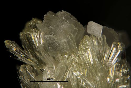
Recent developments in photography have lead to a flood of great images with tremendous sharpness or resolution. Here’s an example photograph of a spherical cluster of blades of vauxite crystals from the Siglo Veinte mine, Llallagua, Potosi, Bolivia. The ball of crystals is 1 mm wide.

This photo was produced by combining the sharpest parts of 55 separate photos of the mineral; each image isolated a tiny, crisply focused level. This photo won an Image of Distinction Award in the 2013 Nikon Small World contest. But how is this done?
Our eyes are a marvelous organs. Light is taken in through the lens of our eyes and this light is focused on the retina. Microscopes or combinations of lenses magnify an image of the microscopic subject. We focus on a portion of the specimen and have the ability to continually focus on different parts of the subject. In producing a photograph, we are limited to one plane of focus and we compromise to record an image of what we want to show in a two-dimensional plane. This compromise is forced because the microscope objective or camera lens system has a fixed depth of field. Unlike a microscope objective, macro lenses on cameras have an aperture diaphragm to adjust depth of field, but that will only help a little. With the proliferation of digital photography, scientists and engineers found a way to take multiple images, focused at different focal planes. The images (image stack – hence “stacking”) are processed using software that selects only the sharpest parts of each photo and produces a composite image with resolution far exceeding the theoretical, physical resolution of the equipment.
Here is a stack of 15 of the photos taken of a vesuvianite specimen with a tiny rhombic calcite impaled on top. The whole specimen is 5 mm wide. The upper left image is focused on the lowest part of the mineral. From left to right, top to bottom, the photos are focused on progressively higher parts of the mineral.
Individually, these are not great photos. Let’s look at what these images looked like to the viewer through the microscope to produce the final image.
For the final image, 28 images were run through a program called Combine ZP (I currently use Zyrene Stacker) software to produce the final crisp image.
In other posts, I discuss the equipment for high magnification, controlling focus stepping, using depth of field to your advantage, and tips to ensure the best image results.

